Nutrition impacts pigmentation, eye migration In Atlantic halibut
Research examines fatty acid composition of enriched artemia, fatty acid ratios
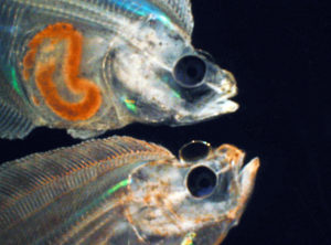
Despite over 15 years of research on Atlantic halibut, a promising aquaculture candidate, its production is still low, partly due to difficulties in juvenile production. The species has a very long yolk sac stage during which it must be kept in the dark for approximately 45 days. Further, juveniles fed artemia in the larval stage often exhibit malpigmentation and lack proper eye migration.
On the other hand, larvae fed their natural diet of copepods under the same conditions show normal development through metamorphosis, showing that the malformations are related to nutrition.
There are a number of differences in nutrient composition between artemia and copepods, but the three factors that have been shown to most affect metamorphosis are fatty acids, vitamin A, and nutrients necessary for the synthesis of thyroid hormone.
Fatty acids
It is generally accepted that high levels of the fatty acids eicosapentaenoic acid (EPA) and docosahexaenoic acid (DHA) are necessary to obtain normal pigmentation in flatfish. Recently, it has also been shown by two research groups that high levels of arachidonic acid (ARA) in live feed produce dramatically reduced pigmentation success in turbot and yellowtail flounders. Contrary to general opinion, research on varying the DHA:EPA ratio showed no effect on pigmentation in turbot.
Data from research at the National Institute of Nutrition and Seafood Research in Bergen, Norway, shows that copepods contain much more EPA and DHA and lower levels of ARA than artemia enriched with an emulsion of phospholipids rich in DHA. The fatty acid composition of the feed is reflected in the larvae.
The 20-carbon fatty acids EPA and ARA are precursors of eicosanoids, which are signalling molecules that regulate a large number of metabolic processes. They compete for the rate-limiting enzyme in eicosanoid synthesis, and the ratio between them determines the ratio of omega-3 and omega-6 eicosanoids in the body. These eicosanoids have different, often counteractive, functions. The malpigmentation of halibut larvae could be a result of low EPA:ARA ratios.
Thyroid hormones
Thyroid hormones are key regulators of metamorphosis in vertebrates. The author and coworkers have looked at the differences in nutrients necessary for the synthesis of thyroid hormone between artemia and copepods and between Atlantic halibut larvae fed the two organisms.
The amino acid phenylalanine is converted to tyrosine, which forms the backbone of the hormones. In the thyroid gland, four iodine are added to tyrosine to form T4, which is later deiodinated to T3, the active form of the hormone. The enzyme responsible for the last step contains selenium.
The author found that phenylalanine, tyrosine, and selenium were more sufficient in artemia than copepods. On the other hand, iodine was 700 times higher in copepods than in artemia and 3-4 times higher in larvae fed copepods compared to larvae fed artemia. T4 was slightly higher in larvae fed copepods during days 15-30 after first feeding, when eye migration is detected.
In another experiment, researchers enriched artemia with iodine to 50 times the level found in control artemia. This led to an increased iodine level and greater tendency of T3 in the Atlantic halibut larvae, but there was no detectable effect on pigmentation and eye migration. Due to suboptimal rearing conditions – large variations in fish size and developmental stages – the experiment will be repeated.
Vitamin A
Japanese researchers have shown that enrichment of live prey with vitamin A stimulates pigmentation in flounders. The problem with this approach is that high vitamin A levels also lead to vertebral deformities in the larvae and juveniles.
Some reports indicate that larvae fed artemia, not copepods, may be vitamin A-deficient. Other reports show toxic levels or forms of vitamin A in the live food. Vitamin A is a key regulator of embryogenesis in vertebrates, where both deficiency and excess of the vitamin lead to malformations in embryos. The effects of vitamin A on flatfish larvae indicate the vitamin is also involved in the regulation of metamorphosis.
Researchers used two high-performance liquid chromatography methods – one detecting retinols, retinal, and retinyl esters; and another detecting retinols, retinal, and retinoic acids – to investigate the vitamin A contents of artemia and copepods. The copepods did not contain detectable levels of vitamin A. Artemia produced peaks with retention times similar to retinoids’, but the ultraviolet-visible spectrums of the peaks were very different from those of retinoids. The study concluded that artemia also contain very little vitamin A.
On the other hand, Atlantic halibut can convert the astaxanthin in copepods and cantaxanthin in artemia to vitamin A. The rates of conversion are such that the carotenoid content in the live feed would supply the vitamin A requirement of the fish. However, larvae fed artemia contained less total vitamin A than larvae fed copepods.
When researchers analysed the different forms of vitamin A, they found no difference in the physiologically active forms retinol and retinal, and the main difference in the storage form, retinyl esters. Nevertheless, larvae fed artemia were able to build up their stores of vitamin A. Artemia therefore probably contain enough carotenoids to supply the vitamin A requirement in Atlantic halibut.
Conclusion
The fatty acid composition of enriched artemia probably provides a good explanation for observed malformations in Atlantic halibut larvae and juveniles. Focus on the causes should be shifted from the DHA:EPA ratio to the concentration of ARA and EPA:ARA ratio. The question as to whether iodine is deficient in artemia should be investigated further.
Marine fish larvae may cover their iodine requirement from the iodine found in seawater. On the other hand, the larvae may have adapted to the high iodine levels found in their natural prey. Excess or deficient vitamin A and toxic forms of the vitamin in artemia are probably not the causes of malformations in Atlantic halibut larvae and juveniles.
(Editor’s Note: This article was originally published in the June 2004 print edition of the Global Aquaculture Advocate.)
Now that you've reached the end of the article ...
… please consider supporting GSA’s mission to advance responsible seafood practices through education, advocacy and third-party assurances. The Advocate aims to document the evolution of responsible seafood practices and share the expansive knowledge of our vast network of contributors.
By becoming a Global Seafood Alliance member, you’re ensuring that all of the pre-competitive work we do through member benefits, resources and events can continue. Individual membership costs just $50 a year.
Not a GSA member? Join us.
Author
-

National Institute of Nutrition and Seafood Research
Bergen, Norway
Related Posts
Aquafeeds
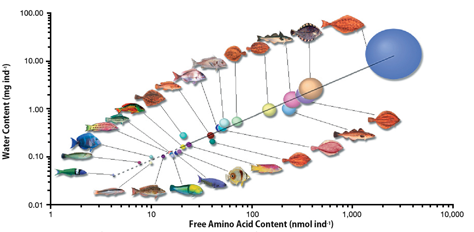
Amino acid requirements in developing marine fish
Most marine fish have pelagic eggs with large pools of free amino acids of fairly constant proportion regardless of species. The amino acids provide energy for metabolism and are an important nutritional asset for growing embryos.
Intelligence
Byproduct utilization for increased profitability, part 4
Protein hydrolysates can be produced by acid, base or enzymatic hydrolysis processes. Acid hydrolysis produces salt that makes the product unsuitable for food and destroys some essential amino acids. An optimum process for one fish or shellfish by-product may not be optimum for another.
Health & Welfare
Enteritis induction by soybean meal in totoaba diets
This study investigated the effects of increasing levels of dietary soybean meal (SBM) with constant taurine supply in the induction of enteritis in juvenile totoaba.
Intelligence
Farmed white halibut
Farmed white halibut initially undergo an 18-month hatchery and nursery stage in land-based tanks. The fish have gained status as a high-quality seafood delicacy.