Southern brown shrimp found susceptible to IMNV
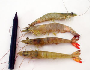
Disease marked by opacity in the abdominal muscles and necrosis of the tails
In September 2002, unusual mortality in farmed Pacific white shrimp (Litopenaeus vannamei) was reported at a shrimp farm in the state of Piaui in northeastern Brazil. Affected animals were lethargic and presented opacity in the second or third abdominal segments. Persistent, low-level mortalities reduced the affected population by 50-70 percent.
Some animals presented reddish coloration in the last abdominal segment and totally necrotic tails. Histopathological analyses showed numerous spheroids in their lymphoid organs and muscular necrosis with hemocytic infiltration. Some specimens presented necrotic lesions in the hepatopancreas.
Infectious myonecrosis virus
Infectious Myonecrosis Virus (IMNV) has since been identified as the causative agent of the necrosis in L. vannamei. Until now, the disease was reported as naturally occurring in cultured L. vannamei. The nonenveloped virus was also experimentally induced in Pacific blue shrimp, L. stylirostris; and black tiger shrimp (Penaeus monodon).
Southern brown shrimp infection
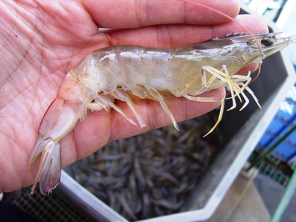
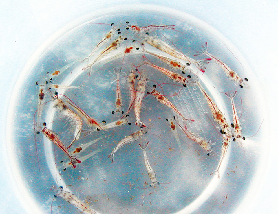
To overcome mortalities produced by IMNV in cultured L. vannamei and increase production, some Brazilian shrimp farmers are testing the southern brown shrimp (Fenneropenaeus subtilis) in grow-out ponds.
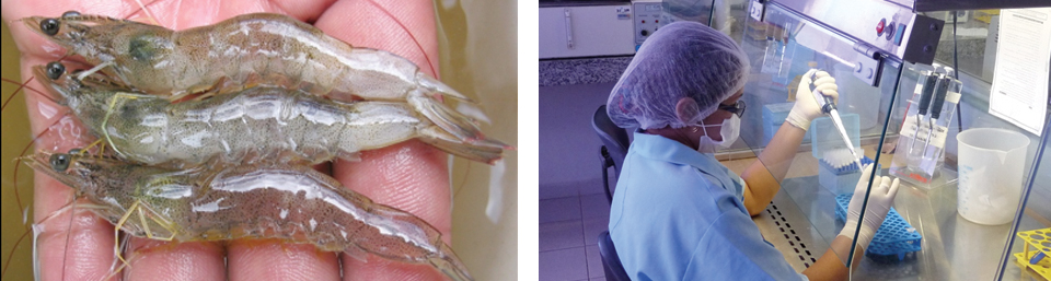
In a recent test at a commercial farm, two 2-ha ponds were stocked with southern brown shrimp at 10 postlarvae per square meter. The animals were fed a 35 percent-crude protein commercial diet formulated for L. vannamei. Dissolved-oxygen levels remained 5-8 ppm during the whole cycle. After 65 days of culture, the first symptoms of IMN were noticed in animals in both ponds. At this time, the shrimp averaged 7 grams in body weight. At harvest, body weights averaged 11 grams, and the survival rates in the ponds were 23 and 26 percent.
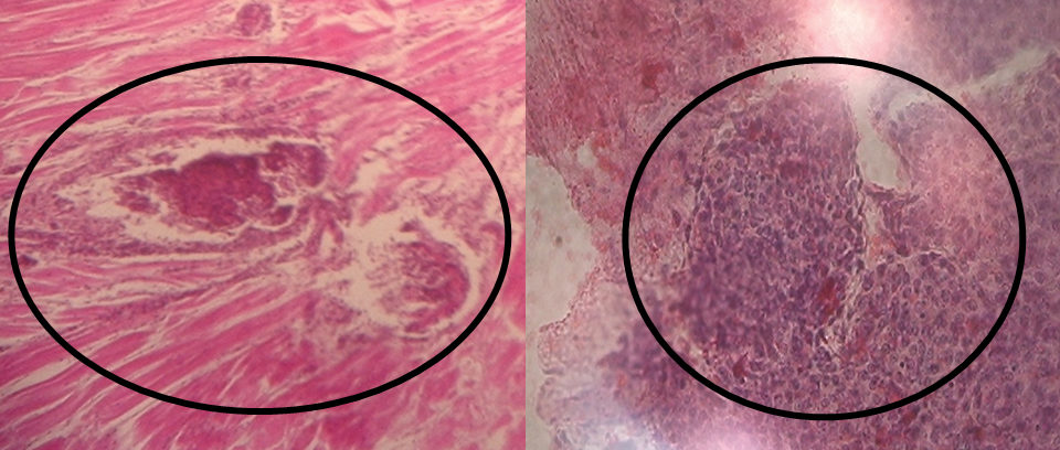
Diseased shrimp presented the characteristic opacity in the abdominal muscles and necrosis of the tails. Histopathological sections stained with the salts hematoxylin and eosin showed spheroids in the lymphoid organs and focal necrosis in muscle, with hemocytic infiltration.
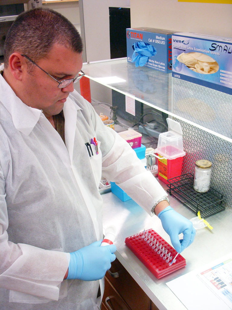
To confirm the diagnosis, reverse transcription polymerase chain reaction (RT-PCR) testing was done using RNA extracted from the gills and pleopods of affected shrimp with a commercial kit for IMNV and primers designed at the University of Arizona. Results confirmed the presence of IMNV in the cultured F. subtilis, and thus expanded the host range susceptibility of IMNV.
(Editor’s Note: This article was originally published in the January/February 2007 print edition of the Global Aquaculture Advocate.)
Now that you've reached the end of the article ...
… please consider supporting GSA’s mission to advance responsible seafood practices through education, advocacy and third-party assurances. The Advocate aims to document the evolution of responsible seafood practices and share the expansive knowledge of our vast network of contributors.
By becoming a Global Seafood Alliance member, you’re ensuring that all of the pre-competitive work we do through member benefits, resources and events can continue. Individual membership costs just $50 a year.
Not a GSA member? Join us.
Authors
-

Centro de Diagnostico de Doenças de Camarão
UnP Campus Salgado Filho
Lagoa Nova, Natal/RN, Brazil -
Centro de Diagnostico de Doenças de Camarão
UnP Campus Salgado Filho
Lagoa Nova, Natal/RN, Brazil -
Instituto de Investigaciones Pesqueras
Facultad de Veterinaria, UDELAR
Tomás Basáñez 1160, CEP: 11300
Montevideo, Uruguay
Related Posts
Health & Welfare
Brazil shrimp farm performs genetic selection for IMNV resistance, growth
The Queiroz Galvão Alimentos shrimp farm and hatchery in Brazil have been working with Concepto Azul to implement a disease-prevention and genetic-breeding program that addresses ongoing impacts from infectious myonecrosis virus (IMNV) and other pathogens.
Health & Welfare
Brazil study: Beta-glucans improve survival of IMNV-infected white shrimp
Authors examined whether beta-glucans extracted from bakers yeast could improve the survival and growth of Pacific white shrimp challenged with infectious myonecrosis virus.
Health & Welfare
Hybrid assay can detect IMNV in resource-poor settings
The method can shorten analysis time and has applications for IMNV diagnosis in resource-poor settings because it does not require specialized equipment.
Health & Welfare
Lab challenge for selection of IMNV-resistant white shrimp
Although still being refined, the IMNV challenge method developed at the University of Arizona has shown promise as a tool to measure resistance in selected family lines of L. vannamei.