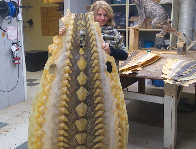
Ultrasound helps stage sturgeons for caviar production
Technique valuable for determining gestational age, fetal viability
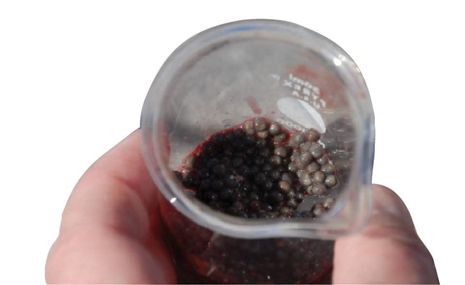
Currently, the only means of sexing sturgeons and measuring oocyte maturity involves biopsying the reproductive tissue of mature sturgeons and measuring its oocyte polarization index (P.I.). P.I. is the measure of the position of the germinal vesicle (nucleus) relative to the animal pole. Initial P.I. measurements are used to determine when during the year the eggs will reach spawning readiness. Generally, optimal caviar yield and quality are reached when the P.I. is 0.10 to 0.12.
Biopsying requires anesthetizing the sturgeons. It is a labor-intensive and invasive procedure in which a 1-cm incision is made along the abdominal wall, and gonad tissue is aspirated. Consequently, this procedure stresses the fish and results in scarring and other undesirable effects.
Due to the time-consuming nature of these procedures, biopsy and P.I. measurements are generally only performed once in the fall to predict the timing of oocyte harvest the following winter and spring. However, P.I. measurements are highly variable among mature females and are not always an accurate predictor of egg maturity, often resulting in improper timing of harvests, decreasing caviar yield and quality, and increasing rates of resorption of eggs.
Additional methods of staging oocyte ripeness are necessary. These methods should be more reliable, non-invasive and, if possible, less time-consuming.
Ultrasound
For many years, ultrasound has been a valuable tool in the medical field and terrestrial animal agriculture for non-invasively assessing soft tissue within live animals. Ultrasound is a cyclic sound pressure with a frequency of roughly 20 kHz or greater. As the wave energy passes through tissues, energy is scattered, absorbed or reflected back to the transducer depending on the properties of the tissue and the relative changes of those properties between tissues.
Ultrasound is most commonly used as a medical diagnostic tool to visualize internal body tissues, such as muscles, tendons and organs. Additionally, ultrasonic imaging (sonography) is a valuable technique for determining gestational age and fetal viability. Ultrasonic imaging offers advantages over magnetic resonance imaging and computed tomography scans that include relative portability, straightforward operation and lower costs.
Oocyte study
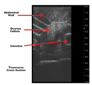
In a study, the authors assessed the utilization of ultrasound in determining both the sex and oocyte maturity in adult white sturgeons. To accomplish this, a portable, waterproof ultrasound machine was used. Transverse and sagittal ultrasonic images were captured at multiple sections along the left and right abdominal walls between the pectoral and pelvic fins of 54 adult female white sturgeons and four adult male white sturgeons.
Among the female fish, 30 were identified as having a distinct, mature egg mass in both the transverse and sagittal planes. These findings were confirmed by biopsy. Furthermore, egg size from each mature female sturgeon was calculated using the measuring tool on the ultrasound device and compared to measurements taken following biopsy, leading to mixed results. The female sturgeons that did not display clear signs of a mature egg mass were found to have immature or atretic eggs, which was also confirmed by biopsy.
Gonad comparison
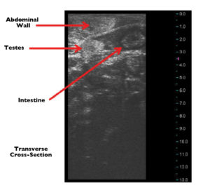
A secondary aspect of this study was to use ultrasonic imaging technology to evaluate the gonad tissue of adult male sturgeons and compare the images to mature female sturgeons. Despite the few adult male sturgeons scanned, the identification of testes was unambiguous. However, the occurrence of large fat deposits made positive identification of testes more difficult than confirming the presence of mature egg masses in adult females.
(Editor’s Note: This article was originally published in the May/June 2010 print edition of the Global Aquaculture Advocate.)
Now that you've reached the end of the article ...
… please consider supporting GSA’s mission to advance responsible seafood practices through education, advocacy and third-party assurances. The Advocate aims to document the evolution of responsible seafood practices and share the expansive knowledge of our vast network of contributors.
By becoming a Global Seafood Alliance member, you’re ensuring that all of the pre-competitive work we do through member benefits, resources and events can continue. Individual membership costs just $50 a year.
Not a GSA member? Join us.
Authors
-

University of Idaho
Hagerman Fish Culture Experiment Station
3059F National Fish Hatchery Road
Hagerman, Idaho 83332 USA -
University of Idaho
Hagerman Fish Culture Experiment Station
3059F National Fish Hatchery Road
Hagerman, Idaho 83332 USA -
University of Idaho
Hagerman Fish Culture Experiment Station
3059F National Fish Hatchery Road
Hagerman, Idaho 83332 USA -
University of Idaho
Hagerman Fish Culture Experiment Station
3059F National Fish Hatchery Road
Hagerman, Idaho 83332 USA -
University of Idaho
Hagerman Fish Culture Experiment Station
3059F National Fish Hatchery Road
Hagerman, Idaho 83332 USA -
University of Idaho
Hagerman Fish Culture Experiment Station
3059F National Fish Hatchery Road
Hagerman, Idaho 83332 USA -
University of Idaho
Hagerman Fish Culture Experiment Station
3059F National Fish Hatchery Road
Hagerman, Idaho 83332 USA -
University of Idaho
Hagerman Fish Culture Experiment Station
3059F National Fish Hatchery Road
Hagerman, Idaho 83332 USA -
University of Idaho
Hagerman Fish Culture Experiment Station
3059F National Fish Hatchery Road
Hagerman, Idaho 83332 USA
Related Posts

Health & Welfare
Chem-free fixes emerging in sea lice saga
Salmon farmers, using emerging technologies, are exploring new methods of sea lice mitigation in an effort to overcome one of the industry’s most persistent problems. New chemical-free innovations show an industry eager to adapt and adopt environmentally safe practices.
Innovation & Investment
Mucosal mapping architect wins aquaculture innovation award
Quantidoc AS in Norway is the commercialization of Prof. Karin Pittman’s years of fish health research. Pittman, winner of this year’s Global Aquaculture Innovation and Leadership Award, utilizes stereology to measure and better understand mucous on gill, gut and skin tissues – the first line of defense for fish.
Intelligence
As sturgeon farming grows, demand concerns emerge
Caviar, or lightly salted sturgeon roe, has been enjoyed for centuries as an expensive gourmet delicacy. After a drastic decline in wild sturgeon stocks, aquaculture stepped in to fill the void. But can farmed supply find lasting balance with market demand?
Health & Welfare
Emerging trends in salmonid RAS, part 1
The benefits realized by recirculating aquaculture systems – including enhanced fish growth, product quality and survival, as well as reduced treatment costs – have made the RAS approach an important production trend.