PCR methods characterize AHPND V.p isolates
Progress in the development of management monitoring strategies for major shrimp disease
This research analyzed the plasmid sequence from the whole genome sequences of AHPND V. parahaemolyticus (Vp) — a serious shrimp disease — isolates and identified a clear geographical variation within the plasmid, and developed PCR methods to characterize AHPND Vp isolates as either Mexico-type or SE Asia-type The finding of a geographic sequence variation in the AHPND virulence plasmid can be used as a marker for monitoring the spread of this disease.
In this study, we analyzed the sequence variations in the large virulence plasmid carrying the pirA- and pirB-like toxin genes, among Vibrio parahaemolyticus isolates causing acute hepatopancreatic necrosis disease (AHPND). We found a variable region (4,243-bp) that corresponds to the geographic collection sites of the AHPND isolates. We then developed and applied a duplex PCR assay that can serve to diagnose AHPND and, further, to distinguish among pathogenic AHPND strains collected from various geographic regions.
Sequence variation of the AHPND virulence plasmid
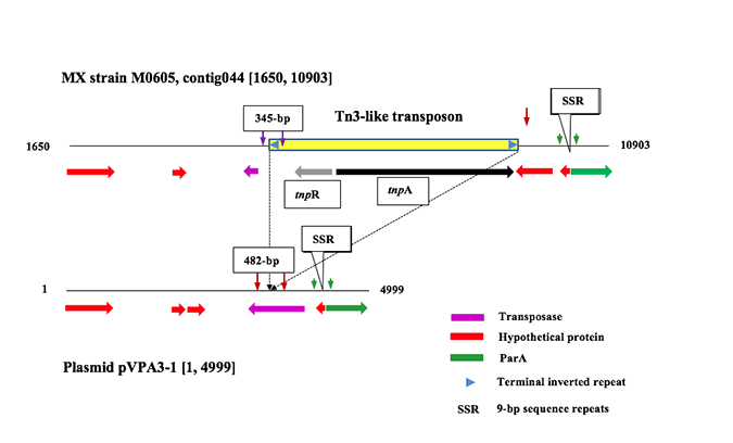
We compared the sequence variation within the virulence plasmid (pVPA3-1) among nine geographic strains (whole genome sequence GenBank no. listed in Table 1). These virulence plasmid’s sequences were identified and assembled from the whole genome sequences (WGS) of AHPND-V. parahaemolyticus by blast analyses. In Mexico AHPND-V. parahaemolyticus strains (M0605 and FIM-S1708+), the Tn3-like transposon is a 4243-bp fragment inserted in the pVPA3-1’s ORF4. However, this fragment is not found in 7 AHPND strains collected from SE Asia including Vietnam, China and Thailand (Table 1, Figure 1).
| Isolates | GenBank No. | Origin (year) | Tn3-like transposon |
|---|---|---|---|
| 13-028/A3 | JOKE00000000 | Vietnam (2013) | - (WGSa & PCRb) |
| NCKU_CV_CHN | JPKU00000000 | China (2010) | - (WGS) |
| NCKU_TV_3HP | JPKS00000000 | Thailand (2013) | - (WGS) |
| NCKU_TV_5HP | JPKT00000000 | Thailand (2013) | - (WGS) |
| TUMSAT_DE1_ S1 | BAVF00000000 | Thailand | - (WGS) |
| TUMSAT_DE1_ S2 | BAVG00000000 | Thailand | - (WGS) |
| TUMSAT_ D06_ S3 | BAVH00000000 | Thailand | - (WGS) |
| M0606 | JALL00000000 | Mexico (2013) | + (WGS) |
| FIM-S1708+ (1 isolate*) | JPLV00000000 | Mexico (2014) | + (WGS & PCR |
| 13-306/D4 (1 isolate*) | NA | Mexico (2013) | + (PCR) |
| 13-511/A1 (1 isolate*) | NA | Mexico (2013) | + (PCR) |
| 14-231 (3 isolates*) | NA | Mexico (2014) | + (PCR) |
| 14-450 (7 isolates*) | NA | Mexico (2014) | + (PCR) |
| 12-194G (1 isolate*) | NA | Vietnam (2012) | - (PCR) |
| 14-188 (5 isolates*) | NA | Vietnam (2014) | - (PCRb) |
| 14-090 (7 isolates*) | NA | Vietnam (2013) | - (PCR) |
| 13-313 (2 samples#) | NA | Country #1 (2013) | + (PCR) |
| 14-433 (3 samples#) | NA | Country #2 (2014) | + (PCR) |
| 15-133 (6 samples#) | NA | Country #2 (2015) | + (PCR) |
| 12-296 (4 samples#) | NA | Vietnam (2012) | - (PCR) |
Tn3-like transposon in Mexico AHPND isolates
To determine if the Tn3-like transposon is only found in the AHPND-V. parahaemolyticus isolates collected in Mexico, we selected a pair of PCR primers MX-345F (5’-TACCAGCTCTAACAAGGCCA) and MX-345R (5’-AACGTTCCAAGGAGTCGAGT) to amplify DNA from AHPND isolates in our collections (Table 1). The forward primer is located upstream of the Tn3-like transposon and reverse primer is located within the Tn3-like transposon (Figure 1). Amplifications were performed with the following parameters: initiation denaturation at 94oC for 3 min, followed by 35 cycles of 94oC for 30 s, 60oC for 30 s and 72oC for 30 s, and a final extension at 72oC for 7 min. We applied this PCR in 29 pure cultures of AHPND-V. parahaemolyticus isolates, and the results showed that all 12 Mexico isolates were positive for the presence of Tn3-like transposon, and none of the 13 Vietnam AHPND isolates were positive for Tn3-transposon (Table 1).
Asian AHPND typing PCR
Another pair of PCR primers Asia-482F (5’-TGAACCGTTCCTCATGCTCT) and Asia-482R (5’-TCAAAGCAGCCCAGACAAAC) selected outside the Tn3-like transposon, within the ORF4 of pVPA3-1, was applied to detect AHPND isolates from Vietnam; the amplicon size is expected to be 482-bp for isolates from SE Asia (Figure 1). The results showed that all 13 pure cultures of AHPND isolates from Vietnam were positive and none of the 13 Mexico isolates were positive. These primers can also anneal to Mexico isolates, but the size of amplicons will be 4732-bp, the PCR products were not detected with the PCR cycling profile used.
Duplex PCR for diagnosing and typing AHPND
For simultaneously detecting AHPND and plasmid typing, 2 pairs of primers — MX-345F/R (or Asia-382F/R) and VpPirA-284F (5’-TGACTATTCTCACGATTGGACTG) / VpPirA-284R (5’- CACGACTAGCGCCATTGTTA) — were added into a single tube during PCR. Amplifications were performed with the cycling profile described above. All of the bacterial cultures showed bands at 284-bp indicating the presence of the toxin gene, as expected. Bacterial cultures isolated from Mexico also showed a band at 345-bp, which indicates the presence of the Tn3-like transposon. In contrast, Vietnam AHPND isolates showed a band at 482-bp, which indicates the absence of the Tn3-like transposon (Figure 2).
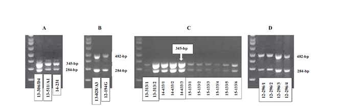
To test this, we applied the duplex method to 15 DNA samples extracted from hepatopancreas tissues of AHPND-affected shrimp. Eleven shrimp were collected from farms in Latin America countries and four were collected from farms in Vietnam. For the Latin American samples, the duplex PCR showed bands at 345-bp and 284-bp (Figure 2), indicating these shrimp were infected with AHPND pathogenic bacteria, as shown by the 284-bp band, and that these bacteria contained a Tn3-like transposon. For the 4 samples collected from Vietnam, the duplex PCR resulted in bands of 482-bp and 284-bp (Figure 2), indicating infection with AHPND bacteria that do not have the Tn3-like transposon. Thus, the duplex method described here is useful for rapid diagnosis and typing of AHPND bacteria of infected shrimp collected from ponds.
Virulence comparison between Mexico and Vietnam isolates
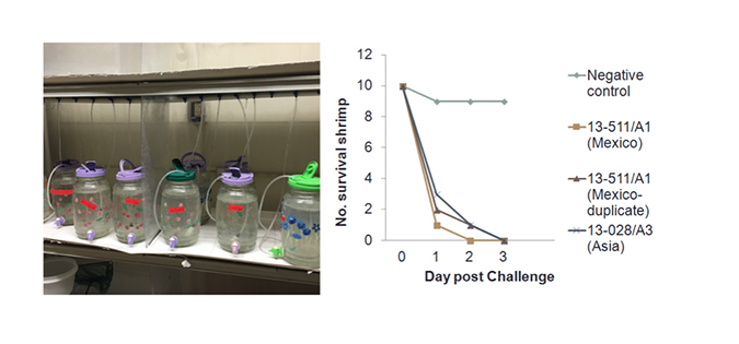
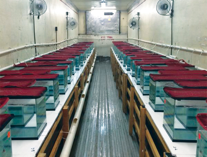
Even though it is found on the virulence plasmid of V. parahaemolyticus, the T3-like transposon appears to be unrelated to the degree of virulence. We have infected P. vannamei with the both Mexico (13-511/A1) and Vietnam (13-028/A3) isolates in the laboratory bioassays. Aquariums (3-L) were filled with artificial seawater at a salinity of 25 ppt and water temperature was maintained at 28°C. Then specific-pathogen free (SPF) P. vannamei (mean weight: 1g) were stocked in the aquariums and fed with V. parahaemolyticus 13-028/A3 and 13-511/A1, respectively, mixed with shrimp feed. The bacteria were grown to 1×10⁹ CFU/mL and mixed with shrimp feed at a 1:1 ratio. The 100 percent cumulative mortality was seen in both isolates by day 3 (Figure 3).
Although the function of the genetic variation among geographic isolates of V. parahaemolyticus remains unknown, the sequence variation such as the presence or absence a transposon can serve as useful markers for detecting the origins of new outbreaks. There are major variation is the presence of a Tn3-like transposon in the Mexico and Latin America isolates that is absent in Asian isolates. It suggests that the Mexico and Latin America isolates share a common origin. In addition, this will aid in the development of management monitoring strategies to limit the spread of the disease and reduce its impact on commercial shrimp farms.
Authors
-

Postdoctoral Research Associate
School of Animal and Comparative Biomedical Sciences, University of Arizona, Tucson, AZ 85721 USA
-

Associate Research Professor
School of Animal and Comparative Biomedical Sciences, University of Arizona, Tucson, AZ 85721 USA
-

Professor Emeritus
School of Animal and Comparative Biomedical Sciences, University of Arizona, Tucson, AZ 85721 USA
Related Posts
Health & Welfare
Genetic variation for resistance to WSS, AHPND in Pacific white shrimp
Selection for disease resistance has been used in breeding farm animals and can be a viable option to deal with white spot syndrome and acute hepatopancreatic necrosis disease in commercial shrimp culture. In trials, heritability for AHPND resistance was low, while that for WSS was moderate.
Health & Welfare
Limited decomposition enhances PCR detection of AHPND Vibrio in shrimp
A study confirmed the utility for improved polymerase chain reaction (PCR) detection of the Vibrio bacteria that cause acute hepatopancreatic necrosis disease (AHPND) in asymptomatic shrimp by permitting the shrimp to expire and decompose for several hours prior to preservation and PCR processing.
Health & Welfare
Measuring susceptibility differences to IHHNV infection
This study evaluated the susceptibility to IHHNV in three different shrimp batches by measuring infectivity titers. Each batch showed a different infectivity titer, therefore, each batch had a specific susceptibility to IHHNV.
Health & Welfare
A holistic management approach to EMS
Early Mortality Syndrome has devastated farmed shrimp in Asia and Latin America. With better understanding of the pathogen and the development and improvement of novel strategies, shrimp farmers are now able to better manage the disease.